
Our Capabilities Include:
- Magnetic resonance imaging (MRI)
- Computed Tomography (CT) scans
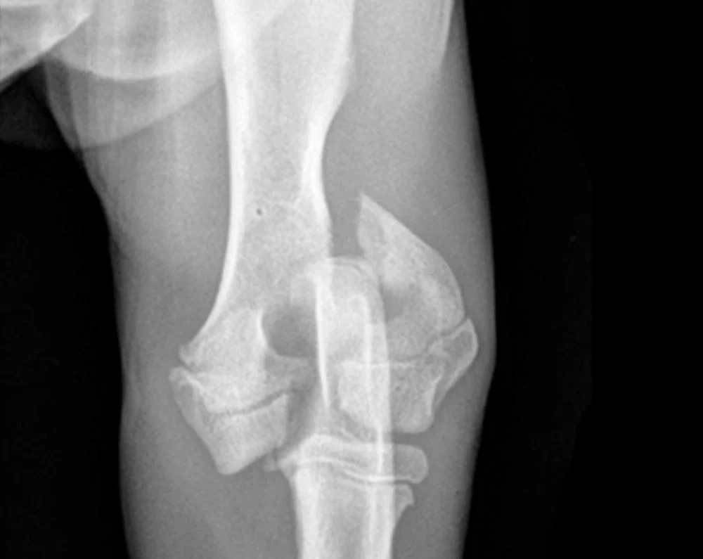
- Digital radiography (X-rays)
- Ultrasonography
- Echocardiography
- Fluoroscopy
What is Advanced Imaging?
Traditional imaging in veterinary medicine has mainly consisted of X-rays. While X-rays are still useful in many situations, MRI, CT, and ultrasound are now available to aid in the diagnostic process. Ultrasound allows for an in-depth look at organs and soft tissues in motion, and Magnetic Resonance Imaging (MRI) and Computed Tomography (CT) scans provide detailed, cross-sectional information about the body in much higher clarity than traditional X-rays.
What’s the Difference Between MRI, CT, Fluoroscopy, and Myelography?
- MRI is safe, non-invasive, and is the ideal method to evaluate the nervous system. It gives us a detailed look at the brain, spinal cord, and nerves. MRI is also an advanced way to look at soft tissue structures such as muscle, ligaments, and tendons. Patients can have MRIs regardless of if they have had implants such as pins, plates, or microchips, but pacemakers and neurostimulators may be contraindicated.
- CT takes X-ray images of “slices” of a patient’s specific organ, muscle, bone, or other internal body part. CT images are much more detailed than a traditional X-ray and can be reconstructed into multiple planes or a three-dimensional model of the affected area, resulting in even greater accuracy. Many conditions which would not be visible using a traditional X-ray can be seen on CT images. CT images are sometimes enhanced with the use of contrast, but MRI is generally preferred for evaluating the nervous system.
- Fluoroscopy uses X-rays to create both images and real-time moving views of the body. It’s used for guiding orthopedic surgery, neurosurgery, interventional procedures, and contrast studies.
- Myelography involves injecting a contrast agent in the space next to the spinal cord and using X-rays to make images of the nervous system. Myelograms used to be the standard method for evaluating the nervous system, however, MRI has now replaced this type of imaging in most situations, due to the fact that MRI gives much more information and is non-invasive. Myelography can still be beneficial in situations where MRI is contraindicated.
Pieper Veterinary offers state-of-the-art diagnostic imaging services for your pets, using the same equipment that you would find at your local human hospital. Different methods of imaging can be used to help diagnose your pet, including digital radiography (X-ray), ultrasonography, echocardiography, fluoroscopy, computed tomography (CT), and magnetic resonance imaging (MRI).
X-ray and abdominal ultrasound (including echocardiography, or cardiac ultrasound) are the most commonly used imaging modalities in veterinary medicine. They are utilized on a daily basis by all primary care doctors, emergency doctors, and specialists at Pieper.
Some Indications for Advanced Imaging Include:
- Cancer screening, staging, or surgical planning
- Neurologic disorders or injuries
- Vascular disease diagnosis and treatment planning
- Traumatic injury assessment
For patients undergoing MRI examination of the nervous system, the neurologist will be able to review the results of the MRI the same day to go over further diagnostic or treatment options. Owners of pets undergoing MRI for areas outside the nervous system will be able to discuss the results of the exam with the clinician who ordered the test.
What is a Radiologist?
Rapid advancements in veterinary care and increased availability of a variety of imaging techniques demand expertise in this area. An American College of Veterinary Radiology (ACVR) board-certified radiologist spends three or more years in a training program dedicated to learning image acquisition and study application and interpretation. Our radiologist works closely with your primary care veterinarian and attending Pieper clinicians to determine what imaging is best suited for the clinical scenario, ensure an accurate and timely diagnosis, and create a plan for further treatment.
Working with your Primary Care Veterinarian
Your general practice veterinarian is an important part of your pet’s health and we work as an extension of their services when advanced or after-hours treatment is necessary. We will notify your veterinarian that your pet visited our diagnostic imaging service, and provide them with treatment and follow-up information to continue your pet’s care once you leave our hospital.

Middletown, CT
Specialty Care
730 Randolph Road
860-347-8387
24/7 Emergency &
Primary Care
Mon–Fri: 8:00 am – 8:00 pm
Sat & Sun: 8:00 am – 4:00 pm
Specialty: By Appointment Only
Meet the Team
Our specialty services team includes experienced veterinarians and skilled veterinary technicians who deliver thoughtful, compassionate care tailored to your pet’s needs.

Bronwen Childs, DVM, MS, DACVR
Veterinarian - Specialist

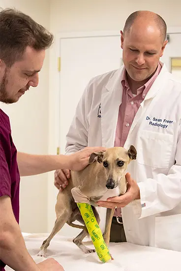
Sean Freer, DVM, DACVR
Veterinarian - Specialist
Sit. Stay. Subscribe.
Monthly pet care tips and updates, minus the fluff.